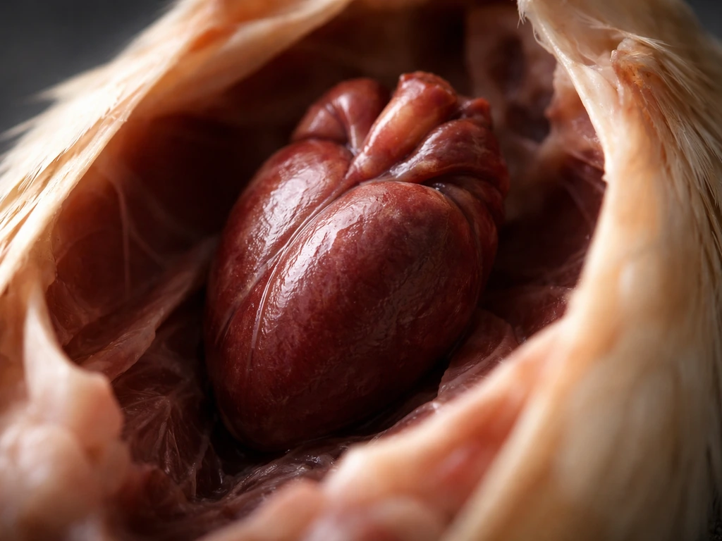
A bird's heart looks like a small, triangular organ sitting upright inside a clear, fluid-filled sac, narrower at the pointed bottom tip and broader at the top where the major blood vessels connect. In fresh specimens it's a deep red to reddish-brown color, noticeably firm and compact, and it sits right behind the breastbone toward the front of the chest cavity rather than deep in the belly. The left side looks slightly bulkier than the right, and the whole thing has that characteristic 'set inside a bag' appearance from the pericardial sac surrounding it.
What Does a Bird Heart Look Like? Size, Shape, and Location

Marcus Whitley
29 Jun 2026
Are you asking about the organ, or a heart-shaped feature?
Before going further, it's worth clarifying what people usually mean when they search for this. Most of the time, someone asking 'what does a bird heart look like' genuinely wants to see the actual cardiac organ, maybe they're looking at an anatomy diagram, watching a dissection video, or studying a necropsy image and need to know which structure is which. That's exactly what most of this article covers.
Occasionally, though, people use 'heart' loosely to mean a heart-shaped mark or region on a bird's body, like a rounded chest patch, a feather pattern, or even a heart-shaped face (the barn owl is a classic example of a bird with a distinctly heart-shaped facial disc). If that's what you're after, the answer is simpler: look for symmetrical, rounded markings or facial shapes in the plumage rather than anything internal. For everyone else curious about the real organ, keep reading.
Bird heart anatomy in plain terms
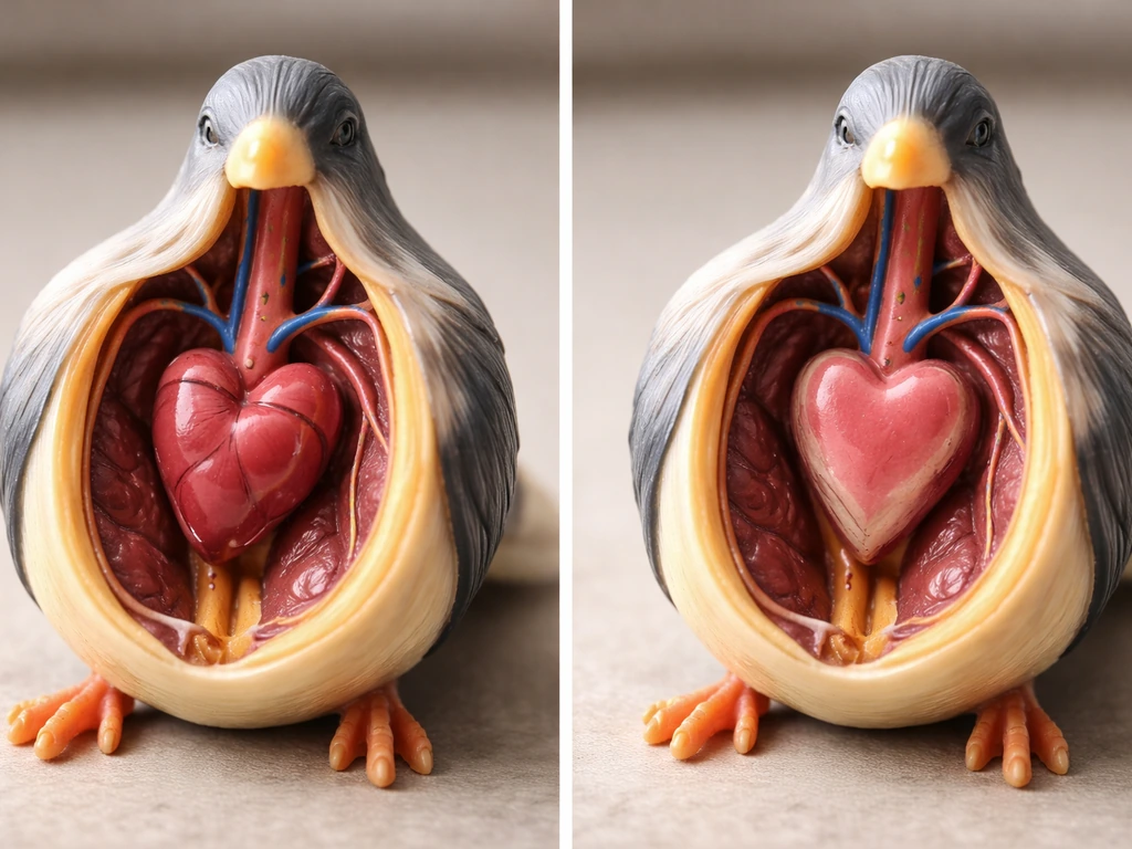
The avian heart works on the same four-chamber plan as a mammal heart, but the proportions look a bit different when you actually see one. The left ventricle is the dominant chamber, thick-walled, cone-shaped, and muscular. It forms most of what you'd call the 'bulk' of the heart and creates that slightly asymmetric, left-heavy appearance you'll notice in diagrams. The right ventricle is crescent-shaped and sits more to the side; it doesn't extend all the way down to the pointed tip of the heart. That tip, the apex, is the caudal-most point of the organ, pointing slightly downward and toward the tail.
The base of the heart, where all the major vessels enter and exit, faces upward toward the bird's head. Think of the heart as a triangle with the point aimed down and the wide end aimed up, sitting almost parallel to the spine and breastbone. That orientation stays consistent across bird species, which makes it a reliable anchor once you know it.
What a healthy bird heart actually looks like up close
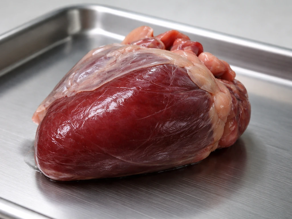
In a fresh specimen, the heart is a deep red to dark reddish-brown, roughly similar to the color of a fresh beef liver but with a firmer, more compact texture. The surface isn't perfectly smooth, you can make out faint grooves where the coronary vessels run, and the boundary between the right and left ventricles is sometimes visible as a subtle groove or fat line on the surface.
The pericardial sac is one of the most useful visual cues. It's a thin, clear to slightly yellowish fibrous sac that surrounds the heart, and inside it there's a small amount of colorless to pale yellow fluid. In fresh or well-preserved specimens, you'll often see the heart sitting visibly inside this sac, almost like a piece of fruit inside a clear bag. That 'bag' is what distinguishes the heart at a glance from surrounding lung tissue or air sacs, which don't have that kind of defined enclosure.
At the base of the heart, where it widens toward the head end, you can trace the major vessels if you look closely, the aorta leaves the left ventricle and curves upward, and the pulmonary vessels are nearby. These vessel roots are a helpful cross-check when you're trying to confirm you've found the right structure.
Where you can actually see a bird heart today
If you're looking for a clear, labeled visual right now, anatomy diagrams are your best starting point. Search specifically for 'avian heart gross anatomy' or 'bird necropsy heart diagram' and you'll find labeled illustrations showing the triangular shape, chamber positions, and surrounding pericardial sac. Veterinary and ornithological textbooks aimed at avian medicine often include the clearest gross-anatomy images.
For hands-on viewing, dissection specimens are the gold standard. In a classroom or lab setting, bird hearts become visible after carefully opening the thoracic cavity and reflecting the sternum. The heart appears in the cranial (front) portion of the chest space, close to the sternum, not buried back near the digestive organs. In larger birds like pheasants or chickens, the heart sits roughly between the first and fourth ribs, a useful mental landmark if you're working with a whole-body specimen. If you’re specifically trying to picture what a calling bird looks like, also consider its size, coloration, and overall body shape rather than its anatomy what does a calling bird look like.
Imaging references (such as radiographs or echocardiograms used in avian veterinary medicine) show the heart as an oval to teardrop-shaped shadow sitting cranioventral in the body cavity, well forward of the stomach and intestines. If you're looking at a bird X-ray, the heart appears as a dense oval structure right behind the keel of the sternum.
How heart appearance changes across bird types
The core look stays the same across species, but scale and relative size shift noticeably. Hummingbirds have hearts that are enormous relative to their body size, proportionally the largest among birds, because their metabolic demands are so extreme. A hummingbird heart is tiny in absolute terms (smaller than a pea) but takes up a remarkable percentage of the bird's chest. At the other end, a large bird like a turkey or emu has a heart roughly the size of a small plum.
| Bird type | Approximate heart size | Notable visual trait |
|---|---|---|
| Hummingbird | Smaller than a pea | Proportionally very large for body size; very fast-contracting |
| Songbird (sparrow/robin size) | Pea to small grape | Classic triangular shape, clearly visible pericardial sac |
| Medium bird (pigeon/crow) | Small grape to marble | Left ventricular bulk visible; vessel roots more defined |
| Large bird (chicken/pheasant) | Marble to small plum | Heart spans roughly from first to fourth rib in dissection |
| Very large bird (turkey/emu) | Small plum or larger | Same triangular orientation; more visible surface coronary grooves |
Regardless of size, the dorsal-to-sternum positioning and the parallel-to-spine orientation hold true across all bird species studied. So whether you're looking at a tiny warbler heart in a diagram or examining a chicken in a lab, the 'where it sits' rules are the same.
Common mix-ups and what preservation does to appearance
The most common beginner mistake is confusing the heart with the air sacs or adjacent lung tissue. Avian air sacs are thin, almost tissue-paper-like membranes that extend throughout much of the body cavity. They look like light, translucent pouches, nothing like the firm, enclosed triangular structure of the heart. If what you're looking at seems flat, membrane-like, or doesn't have a clear sac boundary, it's almost certainly an air sac or pleural tissue, not the heart.
Another mix-up involves the thymus, which in some birds sits in the cranial chest region near the great vessels. The thymus is softer, more lobular, and pale pinkish-gray rather than the deep red of cardiac tissue. It also sits more cranially (closer to the neck/head end) than the heart itself, so if your 'heart' candidate is up near the base of the neck, it's worth double-checking.
Fat pads can also throw beginners off. Pericardial fat, which accumulates around the heart in well-nourished birds, shows up as yellowish, soft tissue partially coating the pericardial sac. It can obscure the clean triangular silhouette you're looking for, but the firm, red organ will still be identifiable once you gently move the fat aside.
Preservation changes color noticeably. Formalin-fixed specimens typically turn the heart a pale grayish-brown or beige rather than the vivid red of fresh tissue. The pericardial sac may also appear more opaque and the fluid inside may be harder to see. Don't let the color shift throw you, focus on the shape (triangular, apex-down), position (cranial, dorsal to sternum), and the sac enclosure rather than trying to match a 'fresh red' color in a preserved specimen.
How to confirm you're actually looking at the heart
If you're working from a diagram, start by checking for labels before anything else. Most anatomical diagrams label the pericardial sac, ventricles, and major vessels, and the heart will almost always be shown in the cranial thoracic region, close to the sternum. If the diagram is unlabeled, locate the sternum first, then look for the triangular, sac-enclosed structure immediately behind and slightly dorsal to it. If you specifically want what a female cat bird looks like, pair those anatomical cues with a reference photo of the species you have in mind so you can compare the heart’s position and shape triangular, sac-enclosed structure.
- Check position first: the heart sits in the front of the chest cavity, near the sternum, not back near the digestive organs.
- Look for the pericardial sac: a clear, bag-like enclosure around the organ is a reliable tell. Lungs and air sacs don't have this wrapper.
- Confirm the triangular shape: base (wider end) toward the head, apex (pointed tip) toward the tail.
- Notice the bulk: the left side should appear slightly more massive than the right, especially toward the apex.
- Trace the vessels: at the base of the heart, look for large vessel roots (aorta and pulmonary vessels). If you can see vessel stumps or roots at the wide end of the organ, you've found the right structure.
- In preserved specimens, rely on shape and position rather than color, since formalin fixation mutes the red coloring significantly.
If you're still uncertain after working through those checks, the single most useful next step is to find a labeled gross anatomy photograph of a bird species close in size to what you're studying and compare side by side. Veterinary resources for avian medicine, wildlife rehabilitation manuals, and comparative anatomy textbooks all include these. The more you compare labeled images to what you're seeing, the faster the heart's distinctive shape and position lock in as something you can spot immediately.
If you find bird internal anatomy interesting, it connects naturally to the broader picture of how birds are built. The heart's position makes more sense, for instance, once you understand how the avian skeleton frames the thoracic cavity, a topic worth exploring alongside this one for anyone wanting the full structural picture of how a bird looks from the inside out. Seeing what a bird skeleton looks like helps you understand how the thoracic cavity is shaped and where the heart will sit avian skeleton.
FAQ
How can I tell a bird heart from the lungs or air sacs in a photo or specimen?
Look for a clearly defined boundary, the heart sits inside a sac (the pericardium) and feels firm. Air sacs tend to be thin, semi-translucent sheets that blend into surrounding tissue without a crisp enclosed “container.”
What changes do I expect in color and visibility between fresh, frozen, and formalin-fixed specimens?
Fresh tissue is typically deep red to reddish-brown, while formalin-fixed hearts often look grayish-brown or beige and the pericardial sac can look more opaque with less visible fluid. In all cases, prioritize the triangular shape (apex-down) and sac enclosure over color matching.
Where exactly should the “apex-down” point be aiming in the bird’s body?
The apex is the lowest point and it points slightly downward, toward the tail end. The heart’s wide base faces upward toward the head side, near where the major vessels connect.
Why does the left side of the heart look bulkier, and is that normal?
The left ventricle is typically the dominant, thick-walled chamber, so it contributes most of the heart’s “bulk.” Mild left-right asymmetry is expected in diagrams and gross anatomy views.
Can the thymus be mistaken for the heart, and how do I reduce that confusion?
Yes, especially in the cranial chest region. The thymus is softer, more lobular, and pale pinkish-gray rather than deep red, and it sits closer to the neck than the heart does. If your “heart” is very near the base of the neck, re-check location.
What should a bird heart look like on an X-ray, and how do I separate it from nearby shadows?
It commonly shows as a dense oval or teardrop-shaped shadow in the cranio-ventral chest region, forward of the stomach and intestines. Use sternum position as your anchor, the heart shadow usually sits just behind the keel rather than deeper in the abdomen.
If I’m looking at a labeled diagram, what landmarks should I use first?
Start with the sternum and the pericardial sac label if present. Then confirm the triangular, sac-enclosed structure immediately cranial to the stomach area, with the apex oriented downward toward the tail.
How can fat around the heart affect identification, and what’s the best way to handle it?
Pericardial fat can blur the clean triangular outline and make the sac less obvious. The heart is still firm and red compared with the softer yellow fat, if you gently shift or tease fat aside you can usually restore the heart’s silhouette.
Do all birds have the same relative heart position, or can it shift by species?
The general “where it sits” rules are consistent, cranial placement behind the breastbone, dorsal-to-sternum orientation, and alignment nearly parallel to the spine. What changes most between species is absolute size and relative proportion, not the core orientation cues.
What’s a practical way to confirm I’m seeing the heart when I only have one reference image?
Compare against a labeled image from a bird of similar size and thoracic proportions. Side-by-side checking of sternum location, sac enclosure, and apex orientation is more reliable than trying to match exact color or texture alone.