In real anatomy diagrams, a bird brain looks like a small, smooth, oval-to-pear-shaped organ, roughly the size of a walnut or smaller depending on the species. It sits nestled inside the skull and lacks the deep wrinkly folds (called gyri and sulci) you'd see on a mammal brain. The surface is mostly smooth, the two hemispheres are visible but not dramatically separated, and the large optic lobes on the sides are one of the most distinctive features you'll spot right away. Bird vision itself can also help you recognize the optic lobes by what they suggest about how birds perceive their world. If you're searching for images and wondering whether what you're looking at is real bird brain anatomy or just a slang meme, this guide will walk you through exactly what to look for.
What Does a Bird Brain Look Like A Visual Guide

What 'bird brain' actually means (anatomy vs slang)
Before you go down a rabbit hole of image searches, it helps to know that 'birdbrain' has two very different meanings floating around online. In common usage, “birdbrain” is primarily US informal slang meaning “a stupid person.” blank" rel="noopener noreferrer">“birdbrain” in common usage. In everyday American slang, blank" rel="noopener noreferrer">birdbrain means a stupid or scatterbrained person. It's listed in Merriam-Webster, Cambridge, and Dictionary.com as an informal insult, and if you browse Reddit discussions about the word, people are almost always using it that way, not talking about actual neuroanatomy. In r/BirdsArentReal discussions, “birdbrain” is commonly framed as slang for calling someone unintelligent, not as a description of bird neuroanatomy using it that way, not talking about actual neuroanatomy.. So if your search results are flooded with memes, jokes, or cartoon images of someone acting foolish, you've landed in slang territory.
The anatomical meaning is a completely different topic. When scientists, veterinarians, or biology students talk about a bird brain, they mean the actual organ inside the skull of birds like pigeons, crows, sparrows, or parrots. And interestingly, that organ is far more sophisticated than the slang gives it credit for. Crows and parrots in particular have brain-to-body ratios comparable to primates, and their neural wiring supports complex problem-solving, tool use, and even long-term memory. The slang reputation is genuinely outdated science.
What a bird brain looks like in pictures and diagrams
When you find a real anatomy image or illustration of a bird brain, the first thing you'll notice is how compact it is. If you're really asking what a bird itself looks like, focus on visible features like size, color, beak shape, and feather patterns what a bird look like. For a common pigeon or starling, the whole brain is roughly the size of a large grape or small walnut. Scaled to their body size it's proportionally significant, but in absolute terms it's tiny. In a photo of a dissected specimen, the organ often appears off-white, pale cream, or light gray, sometimes with a slightly yellowish tint from preservation fluids. Fresh tissue can look more pinkish-gray.
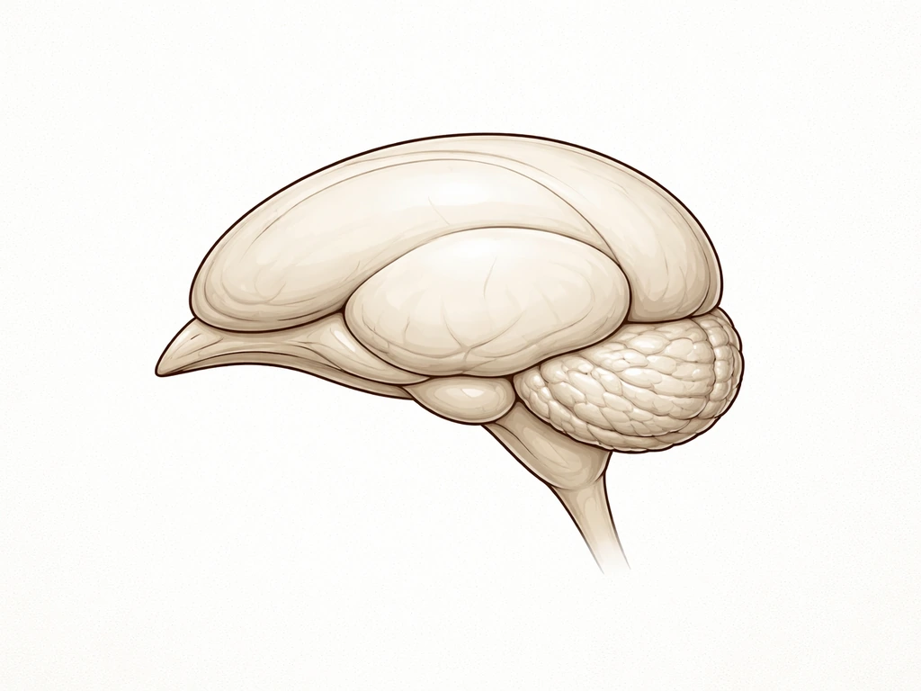
The overall shape is what really sets it apart. Think of a smooth, slightly flattened oval that's widest near the back and tapers toward the front. There are no dramatic ridges or folds on the surface. The two cerebral hemispheres sit on top and are gently rounded, like two soft domes side by side. Below and toward the back, you'll see the cerebellum, which has a finely ridged texture that's easy to spot. On the sides, the optic lobes bulge outward noticeably, and in birds with large eyes (like owls or pigeons), those lobes can be remarkably prominent. In color diagrams, the different regions are usually coded in distinct shades like blue, green, orange, and yellow to help you tell them apart.
Key landmarks to spot in bird brain diagrams
If you're looking at a labeled diagram, here are the major regions you should be able to find. Getting familiar with these makes it much easier to interpret any image you come across.
- Cerebral hemispheres: The large, smooth, rounded domes on top. In birds these take up a big portion of the brain but lack the deep folds seen in mammals.
- Optic lobes (optic tectum): Rounded bulges on the sides of the brain, often quite large. Birds rely heavily on vision, and in many species these lobes are visually dominant in side-view diagrams.
- Cerebellum: Located at the back and bottom, it has a finely layered or ridged surface texture that contrasts with the smooth hemispheres above. It coordinates movement and balance.
- Olfactory bulbs: Small projections at the very front tip of the brain. In most birds these are tiny, though in vultures and kiwis they're proportionally larger.
- Brainstem (medulla): The stalk-like region at the base connecting the brain to the spinal cord. Usually visible in side-view diagrams as a narrowing at the bottom.
- Wulst: A thickened raised ridge on the top of the cerebral hemispheres, visible in some birds and important for sensory processing. Not always labeled in basic diagrams but worth knowing.
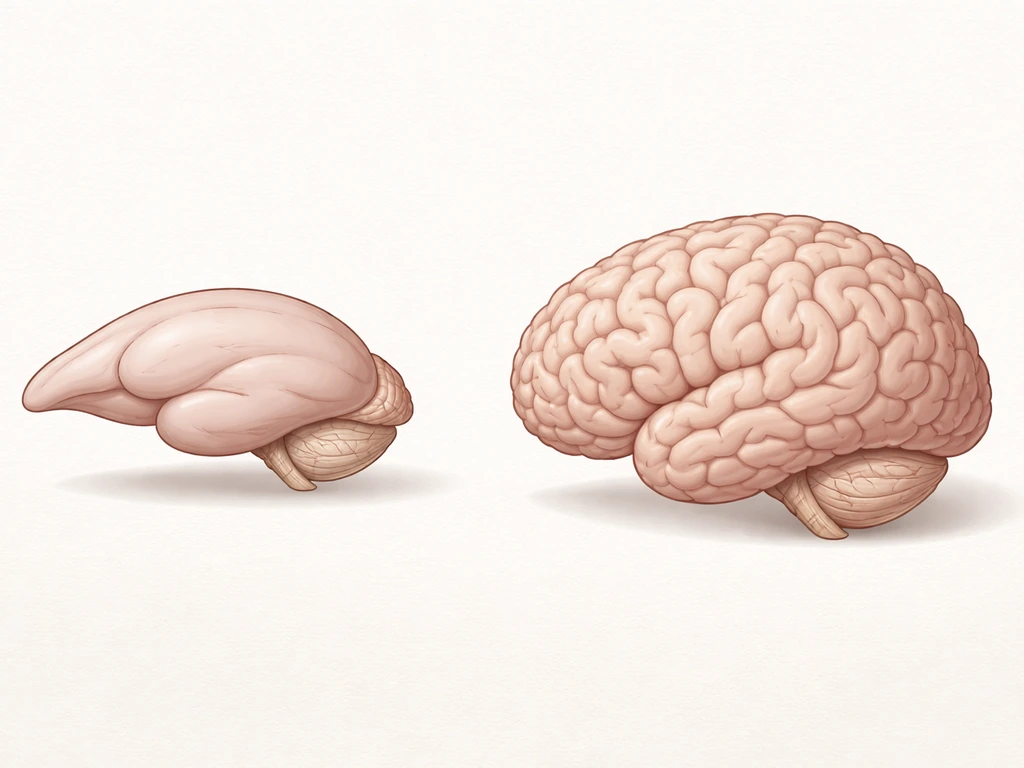
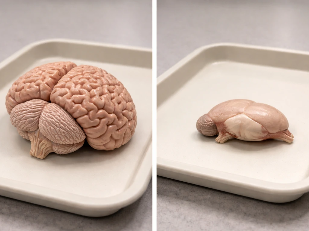
How it compares to a mammal brain visually
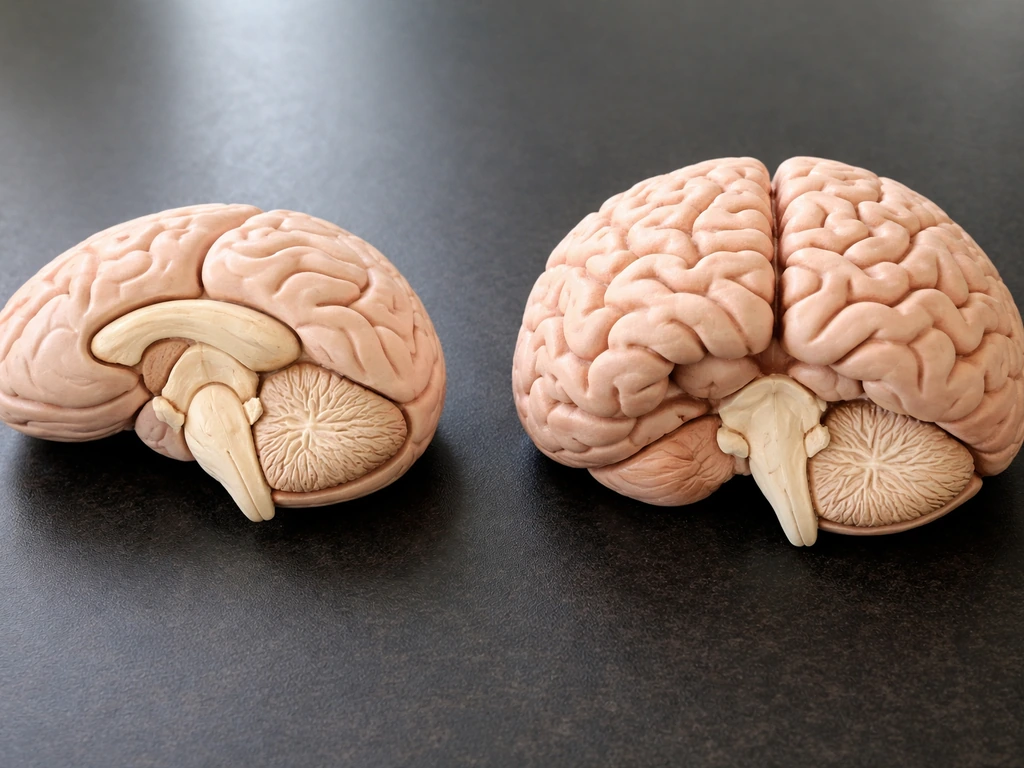
Put a bird brain diagram next to a human or cat brain diagram and the differences jump out immediately. The most striking visual gap is surface texture. Mammal brains, especially primates, are covered in deep folds and convolutions that give them a wrinkled, cauliflower-like appearance. A bird brain surface is smooth and clean by comparison. It almost looks unfinished next to a mammal brain, even though the underlying neural complexity is real.
| Feature | Bird Brain | Mammal Brain (e.g. cat or human) |
|---|---|---|
| Surface texture | Smooth, minimal folding | Deeply folded, wrinkled (gyri and sulci) |
| Overall shape | Compact oval, slightly pear-shaped | Larger, rounder, more elongated in humans |
| Optic lobes | Prominent, visible as side bulges | Smaller, tucked under cerebrum |
| Cerebral hemispheres | Smooth domes, less divided | Clearly divided with prominent folds |
| Olfactory bulbs | Typically small or tiny | Varies; often larger in dogs and cats |
| Cerebellum position | Visible at rear, finely ridged | Tucked under the cerebrum, still ridged |
| Color in preserved specimens | Pale cream, off-white, light gray | Similar pale gray, sometimes slightly darker |
One other difference worth noting: in side-view diagrams, the bird brain's optic lobes often dominate the visual, sitting prominently on the side like two round bumps. In a mammal brain diagram from the same angle, the cerebrum tends to dominate the whole picture, with the optic structures much less visually obvious. That alone is usually enough to tell the two apart at a glance.
Where to find trustworthy bird brain images
Getting your hands on reliable visuals takes a little more effort than a quick image search, but it's worth it. If you are trying to visualize what a bird dog looks like, look for photos from the same angles and lighting across reputable sources so the coat and body type are easy to judge. Here's where to look depending on what you need.
- University anatomy atlases: Many university biology and veterinary departments publish free online atlases of avian anatomy. Search for 'avian brain atlas' or 'pigeon brain diagram' alongside a university name for peer-reviewed, labeled illustrations.
- National Institutes of Health (NIH) databases: The NIH's open-access research portal and PubMed host published neuroscience papers on avian brains, many of which include high-quality labeled diagrams and MRI cross-sections.
- The Cornell Lab of Ornithology: While focused more on bird identification and behavior, their educational materials occasionally include anatomy references and link to credible scientific sources.
- Veterinary anatomy textbooks: Books like 'Avian Medicine and Surgery' or 'Anatomy of Domestic Animals' include detailed bird brain illustrations. Many libraries carry these, and some editions are partially available through Google Books.
- Research articles on crow and parrot intelligence: These species have been heavily studied, and papers on their cognition often include labeled brain diagrams comparing regions across species. Searching 'corvid brain anatomy' or 'parrot telencephalon diagram' gets useful results.
- Natural history museum image libraries: Institutions like the Smithsonian or the Natural History Museum in London sometimes make anatomical illustrations publicly available through their digital collections.
When evaluating any image you find, look for a scale bar or size reference in the photo, labeled regions with standard anatomical names, and a source tied to a published study or academic institution. Illustrations without any labeling or scale reference are often decorative rather than scientifically accurate.
Common mix-ups and how to avoid them
The most common confusion people run into is mistaking the cerebellum for the whole brain. In many dissection photos taken from a certain angle, the finely ridged cerebellum dominates the frame, and it looks quite different from the smooth cerebral hemispheres. If you're only seeing a ridged, layered structure, you're probably looking at the back of the brain, not the whole thing. Step back and find an image with a full side view or a top-down view so you can see all the regions in context.
Another easy trap: using a reptile brain image when you meant to find a bird brain. Reptile and bird brains share some evolutionary heritage and look broadly similar at first glance, both being compact and smooth. The key difference is that bird brains are proportionally larger for their body size and the optic lobes in birds tend to be more pronounced. If the image source is unclear, check whether it's labeled as 'avian' specifically rather than just 'vertebrate' or 'non-mammalian.'
Slang memes are their own category of confusion. If you search 'bird brain' and get cartoon images of a bird with a tiny walnut inside its head, a joke diagram labeled 'bird brain = tiny brain,' or social media posts using the phrase as an insult, those have nothing to do with real anatomy. If you are specifically trying to figure out what a bird chest looks like, use clear photos that show the chest area and the surrounding feathers bird brain. The joke actually gets the science backwards. Modern neuroscience has shown that birds like crows, parrots, and even pigeons have remarkable cognitive abilities, and calling something a birdbrain is more of a compliment than people realize. For the actual anatomy, add the word 'anatomy,' 'diagram,' or 'avian neuroscience' to your search to filter out the noise.
One last mix-up worth mentioning: some people searching this topic are actually curious about a bird's physical appearance more broadly, including things like the chest, eyes, or overall body structure. If that's closer to what you're after, the visual anatomy of a bird's external features like plumage, chest shape, and eye placement is a different but equally fascinating subject. Understanding how a bird's large optic lobes translate into those distinctive wide-set eyes visible from the outside is actually a great bridge between the internal anatomy and the external features you'd notice in the field. If you're trying to picture how a catbird looks, check its typical size, coloring, and distinctive face markings in photos what does a cat bird look like.
FAQ
If I can only find a “bird brain” photo without labels, how can I tell what part I am looking at?
Look for three cues together: smooth front-to-mid brain surface, a ridged structure toward the back (cerebellum), and two prominent side bulges (optic lobes). If you only see ridges and no smooth domes, it is likely a cropped view of the back of the brain rather than the whole organ.
Do bird brains look the same across species, like pigeons versus parrots or crows?
The core layout stays similar, but proportions vary. Birds with very large eyes often show larger, more visually dominant optic lobes, and the overall size can shift noticeably between small passerines and larger corvids or parrots. A diagram for one species can mislead if you expect identical prominence.
Why do some bird brain images look yellowish or gray, and others look more pinkish?
Color often reflects preservation and dissection conditions. Specimens kept in preservative can appear pale cream or light gray, while fresher tissue can look more pinkish-gray. Do not judge “realness” by color alone, use anatomy landmarks like optic lobes and cerebellar ridges.
What search terms help me avoid slang memes when I am trying to find actual anatomy?
Add anatomy qualifiers such as “avian brain diagram,” “bird neuroanatomy,” “pigeon brain dissection,” or “cerebellum optic lobes avian.” If results still show cartoons or “tiny walnut brain” humor, exclude terms like “meme” and “slang” and switch to academic or museum collection wording.
How can I confirm the image is actually avian and not reptile or another vertebrate?
Check for labeling that says “avian” or use anatomical context. A common giveaway is optic lobes that are more pronounced relative to the overall smooth brain shape. If there are no clear labels, rely on the side-view dominance of optic regions and avoid sources that only say “vertebrate brain.”
What’s the quickest way to distinguish the bird cerebellum from the rest of the brain in a side view?
Find the one structure with a finely ridged, layered texture. If that ridged region fills most of the frame, you are likely viewing the back of the brain. A complete side view should show smooth cerebral domes plus the ridged cerebellum, not just one texture.
Are bird brains smaller than mammal brains, and can that affect how images look?
In absolute size, bird brains are typically much smaller than mammal brains, often on the order of walnut or grape scale for common species. Because of that, diagrams may be simplified or enlarged for clarity, so always check for a scale bar or stated specimen size when present.
Do I need to worry about “stylized” illustrations that are not scientifically accurate?
Yes. Decorative diagrams can exaggerate colors or region boundaries. Prefer images with standard anatomical names, consistent region segmentation, and a scale reference. If an illustration has no labels and no indication of species or viewpoint, treat it as educational art rather than anatomy documentation.
If my real goal is understanding what bird brains do, what should I look for beyond visuals?
Search for “avian cognition” or “bird problem-solving neural basis” alongside neuroanatomy terms. Visual guides can tell you where major regions are, but function is usually supported by studies on memory, tool use, and sensory processing that connect those regions to behavior.